The sclera around the optic nerve had more visible blood vessels and was of a different color than the sclera in the immediate vicinity of the cornea. While this could have been the result of our less than surgical removal of the fat and muscle surrounding the area, the fact that (at least in the case of our specimen) the fat and muscle of our eye was concentrated most thickly there suggests that the majority of blood vessels running through the eye enter the sclera in that region.
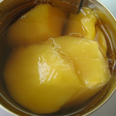
The sclera itself was stiff, and cutting through it with a scalpel was like cutting through posterboard that retained its rigidity when wet. The vitreous humor clung somewhat to the inside of the eyeball. The best simile I could find to describe its texture is that of canned peaches or mangoes. It was moist and slippery, and it was solid but soft to the point of being almost amorphous. However, it could be broken up into distinct chunks when enough pressure was applied. We saw the aqueous humor only briefly, when it rapidly drained out through the pupil following the removal of the (in our case, cloudy, peach- and off-white-colored lens that was hard throughout, like cartilage).
The tapetum displayed iridescence reminiscent of an abalone shell. In the case of our eye, the tapetum seemed not to wholly cover the eye's back, instead being present as an irregularly-shaped blotch with indistinct edges. Over it, the retina hardly seemed like a cohesive layer of cells. It stayed in place despite the absence of the vitreous humor, being possibly stickier than normal due to slight decay, and was torn and punctured so easily that it was more like coating of slime than the membrane I imagined. Opposite the retina, the iris, with the lens removed, resembled the gills of a mushroom with the stem removed. It consisted of two rings of narrow striations. The cornea indeed made noises when stabbed with a scalpel; it was, to add another unnecessary food simile, like a moist, floppy, but still crunchy piece of iceberg lettuce.
What piqued my interest most were the attached muscle and fat. On our eye, it was difficult to distinguish the two from each other, by sight or by touch, much less discern individual muscles or where they attached since everything stuck together. If anything, I'd like to see a cross-section of the entire eye socket to determine how the eye fits in with all the tissues and organs surrounding it.
No comments:
Post a Comment